
Hip Dysplasia in Adults: Why It’s Missed, What It Feels Like, and What Evidence Says Helps
- Emma Glynn
- Feb 23
- 5 min read

By Emma Glynn - The Hip & Knee Physio

What is hip dysplasia?
Hip dysplasia occurs when the hip socket (acetabulum) does not fully cover the femoral head. Instead of a deep, stable “cup,” the socket is relatively shallow, meaning load is distributed over a smaller surface area.
In infants, this is screened routinely. In adults, however, mild or borderline dysplasia is often missed until symptoms develop later in life (Clohisy et al., 2009).
It is not simply a “loose hip.” It is a structural variation that may increase joint stress and labral loading over time.
What does adult hip dysplasia feel like?
Common patterns include:
Deep groin pain
Activity-related ache with walking or stairs
Fatigue in the hip with longer activity
Clicking, catching, or instability
Difficulty with single-leg tasks
Pain that worsens after prolonged standing
Some people describe a feeling of “giving way” rather than sharp pinching (which is more typical of FAI).
Symptoms may overlap with labral tears, hip OA, or gluteal tendinopathy.
Why is dysplasia often diagnosed late?
There are three key reasons:
1. Symptoms mimic other hip conditions
Dysplasia can look like labral pathology or tendon overload.
2. Many adults have never been screened
Mild dysplasia may not be detected in childhood.
3. There is no single “physio test”
Suspicion comes from history + clinical exam + imaging, particularly X-ray measurements such as the lateral centre-edge angle (Wyles et al., 2017).
How is hip dysplasia diagnosed?
Clinical assessment
A physiotherapist may notice:
Reduced hip stability under load
Pain in deep flexion
Muscle fatigue in abductors
Instability symptoms
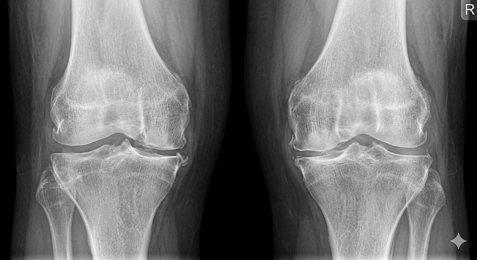
Imaging
X-ray is the primary imaging modality. It evaluates:
Socket depth
Coverage of femoral head
Acetabular angle
MRI may assess labral damage but does not diagnose dysplasia alone.
Importantly, structural variation does not always equal pain. Many people with borderline dysplasia are asymptomatic (Clohisy et al., 2009).
What happens biomechanically?
Because the socket is shallower:
The labrum carries more load
Cartilage stress increases
Hip abductors must work harder for stability
Over time, this may contribute to labral tears or early osteoarthritis (Ganz et al., 2008).
However, progression varies widely between individuals.
Hip dysplasia vs FAI vs hip OA
Feature | Dysplasia | FAI | Hip OA |
Socket depth | Shallow | Normal/deep | Often normal |
Instability feeling | Common | Rare | Rare |
Pinching in deep flexion | Sometimes | Common | Possible |
Age of onset | Young–mid adult | Young–mid adult | 45+ typical |
Risk of early OA | Increased | Increased | Already degenerative |
Does hip dysplasia always require surgery?
No.
Surgery (such as periacetabular osteotomy) is considered in selected cases with significant structural deficiency and ongoing symptoms (Wyles et al., 2017).
However, many adults with mild or borderline dysplasia are managed conservatively first.
What does evidence-based rehab focus on?
While research specific to dysplasia rehab is limited, principles from hip stability literature apply.
1. Improve dynamic stability
Gluteus medius and gluteus maximus strength
Deep rotator control
Pelvic control during single-leg loading
2. Load management
Avoid aggressive end-range hip positions early (deep flexion + internal rotation).
3. Avoid excessive stretching
Over-stretching may increase joint instability.
4. Gradual exposure to activity
Walking, gym, and sport are progressed based on tolerance — not fixed timelines.
Rehab does not “reshape” the socket. It improves how the joint tolerates load.
Who Is at Risk of Adult Hip Dysplasia?
Hip dysplasia is not random. Several factors are associated with increased likelihood of symptomatic dysplasia later in life.
1. Female sex
Hip dysplasia is significantly more common in females. Anatomical and hormonal differences are thought to contribute, including ligamentous laxity and pelvic morphology (Clohisy et al., 2009).
Women are also more likely to present with symptomatic labral tears secondary to dysplasia.
2. Family history
There is evidence of familial clustering. If a parent or sibling had hip dysplasia, early hip arthritis, or required hip surgery at a young age, risk may be higher (Wyles et al., 2017).
3. History of childhood hip issues
Even if you were never formally diagnosed, history of:
Hip braces
Delayed walking
Breech birth
Clicking hips as a baby
may increase suspicion.
Many mild cases go undetected in infancy.
4. High-impact sport participation
Repetitive pivoting, running, and cutting sports may not cause dysplasia but they can expose underlying structural vulnerability, especially if socket coverage is already borderline.
Examples:
Netball
AFL
Dance
Gymnastics
5. Generalised joint laxity
People with hypermobility (e.g., Beighton score elevated) may experience greater instability if acetabular coverage is shallow.
However, laxity alone does not equal dysplasia.
6. Co-existing structural variations
Hip dysplasia often overlaps with:
Labral tears
Borderline acetabular coverage
Femoral version differences
The combination can influence symptom onset and progression.
Does Having Risk Factors Mean You’ll Develop Arthritis?
Not necessarily.
While dysplasia increases relative risk of earlier hip osteoarthritis, progression varies significantly between individuals (Ganz et al., 2008).
Key modifiers include:
Muscle strength
Activity load management
Body mass
Injury history
Structure influences risk. It does not determine destiny.
When should you get assessed?
Consider booking an assessment if:
Groin pain persists beyond 2–3 weeks
You feel instability or giving way
You have recurrent flare-ups
You’re unsure whether symptoms are tendon, labrum, or joint driven
You’re considering imaging or surgical consultation
What you can do this week
Avoid deep loaded flexion temporarily
Reduce repetitive pivoting
Begin controlled hip abductor strengthening
Monitor pain response over 24 hours
If symptoms escalate or include severe night pain, seek individual review.
Internal Links
Can't Find What You're Looking For?
Explore more of our most popular services and guides:
Disclaimer
The content provided on this website is for general information and educational purposes only. It is not a substitute for professional medical advice, diagnosis, or treatment.
While The Hip and Knee Physio strives to present accurate and up-to-date information, we do not guarantee results or outcomes based on the information provided. Any exercises, strategies, or recommendations featured on this site should not be considered a personalised treatment plan.
Always seek the advice of a qualified healthcare provider before starting any exercise program, particularly if you are experiencing pain, injury, or a pre-existing medical condition.
Use of this website does not create a physiotherapist–patient relationship. The Hip and Knee Physio accepts no responsibility for any injury or loss arising from reliance on or use of this information.
By using this website, you agree to these terms.
FAQs
Can hip dysplasia cause back pain?
It may contribute to altered pelvic mechanics, which can influence lumbar load, though this varies individually.
Is dysplasia genetic?
There is evidence of familial clustering, but multiple factors influence development (Clohisy et al., 2009).
Can it get worse with age?
It may increase risk of labral injury and osteoarthritis in some individuals (Ganz et al., 2008).
Do I need surgery?
Not always. Many cases begin with structured rehabilitation.
Can exercise make it worse?
Poorly selected or high-compression movements may aggravate symptoms. Appropriate loading is usually beneficial.
References
Clohisy, J. C., Baca, G., Beaulé, P. E., et al. (2009). Descriptive epidemiology of femoroacetabular impingement: A North American cohort. American Journal of Sports Medicine, 37(Suppl 1), 64S–73S.
Ganz, R., Leunig, M., Leunig-Ganz, K., & Harris, W. H. (2008). The etiology of osteoarthritis of the hip. Clinical Orthopaedics and Related Research, 466(2), 264–272.
Wyles, C. C., Heidenreich, M. J., Jeng, J., et al. (2017). The John Charnley Award: Redefining the natural history of osteoarthritis in patients with hip dysplasia. Clinical Orthopaedics and Related Research, 475(2), 336–350.
(2016). "Imaging of Hip Pain: From Radiography to Cross-Sectional Imaging Techniques". Radiology Research and Practice. 2016: 1–15. DOI:10.1155/2016/6369237. ISSN 2090-1941. Attribution 4.0 International license (CC BY 4.0)
Comments